400-621-6806

2023-06-14 Hits(381)
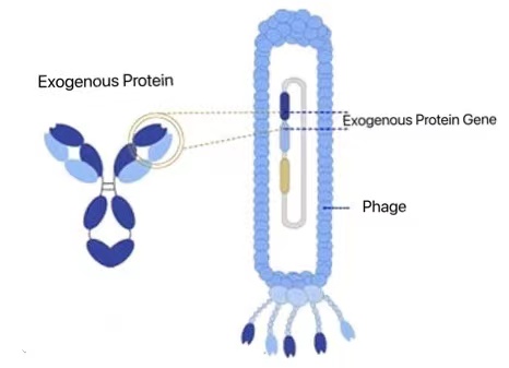
Phage library is a phage carrier used for gene cloning. Phage display library is a new technology that fuses foreign proteins or polypeptides with phage coat proteins, displays them on the surface of phage and maintains a specific spatial conformation, and uses specific affinity to screen specific proteins or polypeptides.
KMD Bioscience has been studied in the field of antibodies for many years. The phage display technology service we have constructed covers a variety of library types, including alpaca/camel VHH antibody library, human antibody phage library, human scFV antibody library, human Fab antibody library, phage peptide library, prefabricated peptide library and other high-quality antibody libraries, which can provide customized services according to customers' needs and provide flexible and effective screening services.
Phage display library technology is a new biotechnology. The foreign proteins obtained by phage display technology maintain independent spatial structure and biological activity. At present, it has been widely used in the fields of new vaccine development, enzyme inhibitor screening, medical diagnosis and treatment, polypeptide drug development, epitope analysis, monoclonal antibody screening, protein interaction research and so on, and shows a good application prospect.
(1) The screening capacity is large, it can be fermented and produced in large quantities, and the method is simple. At present, the storage capacity of the phage primary immune library we obtained can reach 108-109, and its sequence abundance is more than 99%, which ensures that the affinity of antibodies obtained by direct screening can reach nM or even pM level.
(2) It is a new screening technology to combine genotype with phenotype, molecular binding activity with bacteriophage expandability.
(3) Rapid production of soluble antigen, combined with the company's existing recombinant protein expression system, can prepare soluble antigen protein with high purity and high biological activity in a short time to support the immunization and screening of phage antibodies.
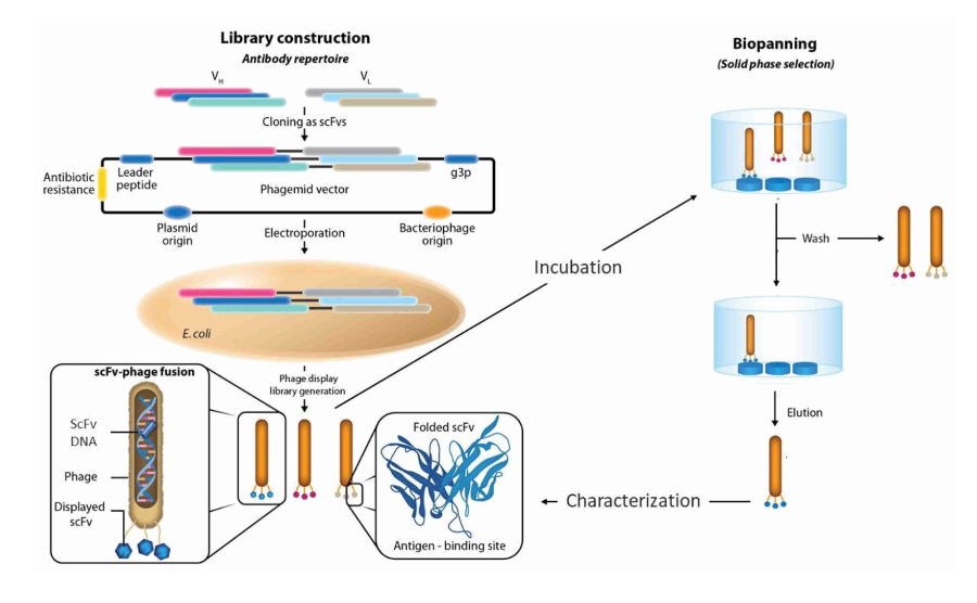
M13 single-stranded filamentous phage display system is a widely used library display system at present. By fusing and expressing proteins, antibodies or polypeptides with phage pIII protein, it can participate in phage assembly and display on the surface of phage, forming a phage display library. KMD Bioscience can construct natural or immune phage antibody display libraries of different species for customers by using M13 filamentous phage shell protein pIII and PVIII display technology. Multi-species monoclonal antibodies, such as those from human, mouse, rabbit, chicken, sheep, goose, pig, cow, horse, donkey, camel, alpaca and shark, are prepared by antibody library technology, which can provide customized services according to customers' needs, and the library capacity can reach 1011.
KMD Bioscience provides customers with phage library construction services, and the process includes: total RNA extraction, reverse transcription to obtain cDNA, PCR amplification, restriction enzyme connection between vectors and PCR products, bacterial library construction, phage library construction and phage library titer detection.
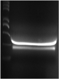
Take the peripheral blood lymphocytes out of the refrigerator, pack them, add chloroform, centrifuge the supernatant, add isopropanol, centrifuge to keep the precipitate, add 75% ethanol, centrifuge to keep the precipitate, add DEPC water after drying, incubate to ensure RNA dissolution, mix all tubes into one tube to obtain total RNA, take 1μl of total RNA for electrophoresis, and take 2μl to measure the concentration with a nucleic acid concentration meter.
According to the operating instructions of the commercial kit, the cDNA was prepared, and the RNA obtained in the last step was divided into two parts and reverse transcribed into cDNA. Oligo dTprimer and random 6-mers were used as reverse primers respectively.
The first round of PCR reaction was carried out with cDNA as template, and PCR amplification was carried out with HS Ex Taq enzyme. The PCR reaction system is as follows:
|
reagent |
application amount |
|
cDNA |
0.5-5μl |
|
AlpVh-L/CALL 002 |
2μl/2μl |
|
dNTP Mix |
4μl |
|
10×ExTaq Buffer |
5μl |
|
HS Ex Taq |
0.25μl |
|
ddH2O |
Up to 50μl |
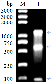
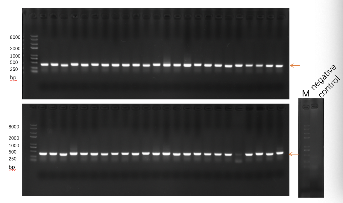
Carry out 1% agarose gel electrophoresis on the PCR amplification products, find the target band to carry out the above conditional PCR amplification, carry out 1% agarose gel electrophoresis on all PCR producing areas, and use the universal DNA purification and recovery kit to recover the DNA from the rubber strips.
The DNA fragments amplified and recovered by PCR in the previous step are used as templates to amplify specific antibody fragments again, and the PCR reaction system is as follows:
|
reagent |
application amount |
|
Recovered products of the first round of PCR |
0.1-2μl |
|
Rvhh FP/RvhhRP |
2μl/2μl |
|
dNTP Mix |
4μl |
|
10×ExTaq Buffer |
5μl |
|
HS Ex Taq |
0.25μl |
|
ddH2O |
Up to 50μl |
The PCR amplification products were recovered according to the instructions by using DNA purification and recovery kit.
The products of the second round of PCR were ligated into the phage plasmid pADL-10b by enzyme digestion, thus constructing a phage plasmid library containing amplified fragments. The ligation products were recovered by DNA purification and recovery kit according to the instructions, and the concentration of the recovered products was detected by nucleic acid concentration measuring instrument.
After the ligation product was transformed by electric shock, an Escherichia coli library containing the target antibody fragment was constructed.
Take the competent cells of SS320 E.coli, add the recovered ligation products, transfer the mixed competent cells and ligation products to the precooled electric rotor, and use the transformation program preset by the electric rotor for electric shock transformation. Immediately after the electric rotor, add 950μlSOC culture medium to the electric rotor for at least 20 electric rotations. After the cells were resuscitated, they were coated on agarose culture plates containing ampicillin resistance for overnight growth. The cells on the culture plate grown overnight in the previous step were washed and scraped off with 2xYT culture medium and a coating rod, and the OD600nm value was measured by adding 20% glycerol and then stored at -80℃, which was the bacterial library.
Mix the bacteria scraped in the last step, then transfer to 100 ml 2x YT culture solution with tetracycline added in advance, and culture at 37℃ and 250rpm until the OD600nm reaches 0.5-0.55.
The auxiliary phage was added according to the ratio of auxiliary phage to bacterial cells of 20:1, and then the culture was continued at 37℃ for 30 minutes. Kana with a final concentration of 50µg/ml and PTG with a final concentration of 0.2mMI were added, and they were cultured overnight in a shaker at 30℃.
The bacteria cultured overnight were centrifuged at 4℃ and 4,000 rpm for 20 minutes. After transferring the supernatant to a new centrifuge tube, 1/4 volume of precooled 20% PEG/2.5m NaCl was added and incubated on ice for 30 minutes. Centrifuge at 4℃ and 4000 rpm for 20 minutes to remove supernatant, and then add 1 ml PBS buffer to dissolve the precipitate. Adding 1/4 volume of precooled 20% PEG/2.5m NaCl again, incubating on ice for 10 minutes, centrifuging at 4℃ for 10 minutes, removing supernatant and dissolving precipitate in 1 ml PBS to obtain phage library, which can be stored at -80℃ for a long time and stored at 4℃ for a short time (1-2 weeks).
After the SS320 strain stored in the refrigerator at -80℃ was cultured, a single colony was selected from the single colony plate and cultured overnight in 5ml 2×YT medium, and the OD600 of the overnight culture liquid was 0.5-0.55. The prepared phage library was diluted in 12 gradients, and 90μl of SS320 bacteria solution was added to each gradient for culture, and 5μl of each dilution tube was added to 2×YT solid medium (Amp) for overnight culture to calculate the titer.
FL-4, Building A5, International Enterprise Community, Tianjin, China
Email:[email protected]
Hot Line: +86-022-82164980
Technical Support:[email protected]
