400-621-6806

Service Line:+86-022-82164980
Address:FL-4, Building A5, International Enterprise Community, Tianjin, China
Email:[email protected]
Introduction
In recent years, Immunology Oncology Therapy has become one of the important means of treatment for advanced malignant tumors. Tumor immunotherapy does not directly attack cancer cells, but by activating the body's own immune system to fight against tumors, with good safety and tolerance. PD-1/PD-L1 antibody drugs, as representative drugs of tumor immunotherapy, have achieved great success in the treatment of advanced malignant tumors.
PD-1, the full name of programmed death Receptor-1, belongs to the CD28 superfamily and is a type I transmembrane protein composed of 268 amino acids. It was discovered in 1992 by Professor Tasuku Honjo of Kyoto University in Japan, and can be expressed on the surface of immune cells such as T cells and B cells. However, when T cells are not activated, PD-1 is almost not expressed, and only after T cells are activated, PD-1 is expressed on the surface of T cells.
PD-L1 was first discovered by Chinese scholar Professor Zhan Ping in 1999 as the third member of the B7 family B7-H1. The following year, Tasuku Honjo and Gordon Freeman of Harvard Medical School demonstrated that B7-H1 can bind to PD-1, inhibit T cell proliferation and cytokine secretion, and negatively regulate lymphocyte activation. Subsequently, B7-H1 was also renamed Programmed death receptor ligand-1 (PD-L1). In addition to being expressed on the tumor surface and participating in immune escape, PD-L1 is also expressed on the surface of antigen-presenting cells (DC cells, macrophages, etc.) and vascular endothelial cells under the stimulation of IFN-γ.
As mentioned earlier, PD-1 can bind to PD-L1, and the two are mutual receptors and ligands. But studies have shown that the relationship is not one-to-one. In addition to PD-L1, PD-1 can also bind to PD-L2. PD-L2 also belongs to the B7 family. Like PD-L1, it is mainly expressed in antigen-presenting cells (DC cells, macrophages, etc.). After binding with PD-1, it can inhibit T cell proliferation and cytokine production, which is related to immune tolerance. Similarly, PD-L1 has been found to bind to other receptors, but is still being explored.
The programmed cell death protein 1 (PD-1) pathway has emerged as a critical regulator of immune responses, playing a pivotal role in cancer immunotherapy. PD-1, a checkpoint receptor expressed on immune cells, interacts with its ligands (PD-L1 and PD-L2) to suppress T-cell activation, allowing tumors to evade immune surveillance. The development of PD-1 antibodies (anti-PD-1 antibodies) has revolutionized cancer treatment by reinvigorating the immune system to attack tumors.
.png)
Figure 1:Immune checkpoint PD-1/PD-L1 signaling pathway [2].
The PD-1 pathway has transformed oncology, with anti-PD-1 antibodies offering unprecedented survival benefits in previously untreatable cancers. As research advances, combination strategies and next-gen inhibitors promise to expand immunotherapy’s reach. With a booming market and ongoing innovations, PD-1 blockade remains a cornerstone of modern cancer treatment, shaping the future of precision medicine.
The PD-1 Pathway: Mechanism and Function
(1) PD-1 Structure and Expression
PD-1 (CD279) is a transmembrane protein belonging to the CD28/CTLA-4 immunoglobulin superfamily. Expressed on activated T cells, B cells, natural killer (NK) cells, and myeloid cells. Its ligands, PD-L1 (B7-H1) and PD-L2 (B7-DC), are found on tumor cells and antigen-presenting cells (APCs).
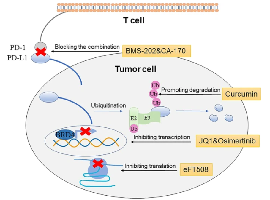
Figure 2:Methods for inhibiting PD-1/PD-L1 signaling pathway [2].
(2) Immune Checkpoint Function
Under normal conditions, the PD-1 pathway prevents autoimmunity by inhibiting excessive T-cell activation. In cancer, tumors overexpress PD-L1/PD-L2, leading to T-cell exhaustion and immune evasion. Blocking PD-1/PD-L1 interaction with anti-PD-1 antibodies restores T-cell function, enabling tumor destruction.
Applications of the PD-1 Pathway
(1) Mechanism of Anti-PD-1 Therapy
PD-1 antibodies (e.g., pembrolizumab, nivolumab) bind to PD-1, preventing PD-L1/PD-L2 engagement. This releases the "brakes" on T cells, enhancing anti-tumor immune responses. Leads to durable responses in multiple cancers, including melanoma, lung cancer, and Hodgkin’s lymphoma[3].
(2) FDA-Approved PD-1 Inhibitors
Several anti-PD-1 antibodies have been approved by the FDA:
|
Drug (Generic Name) |
Brand Name |
Approved Indications |
|
Pembrolizumab |
Keytruda® |
Melanoma, NSCLC, HNSCC, Hodgkin’s lymphoma, gastric cancer |
|
Nivolumab |
Opdivo® |
Melanoma, NSCLC, RCC, HCC, urothelial carcinoma |
|
Cemiplimab |
Libtayo® |
Cutaneous squamous cell carcinoma, NSCLC |
|
Dostarlimab |
Jemperli® |
Mismatch repair-deficient (dMMR) endometrial cancer |
These drugs have demonstrated remarkable efficacy, with some patients achieving long-term remission.
Market Analysis and Future Prospects
(1) Current Market Landscape
The global PD-1/PD-L1 inhibitor market was valued at $35 billion in 2023 and is projected to exceed $60 billion by 2030.
Key players: Merck (Keytruda), Bristol-Myers Squibb (Opdivo), Regeneron (Libtayo).
Keytruda (pembrolizumab) dominates with over $25 billion in annual sales, making it the top-selling immunotherapy drug.
(2) Future Directions
Combination Therapies: PD-1 inhibitors are being tested with CTLA-4 inhibitors (ipilimumab), chemotherapy, and targeted therapies.
Biomarker Development: PD-L1 expression and tumor mutational burden (TMB) are being refined to improve patient selection.
Expansion to New Cancers: Ongoing trials explore anti-PD-1 antibodies in glioblastoma, pancreatic cancer, and rare tumors.
Next-Gen Checkpoint Inhibitors: Bispecific antibodies (e.g., PD-1/TIM-3, PD-1/LAG-3) aim to overcome resistance.
FAQs
Q1: How do PD-1 antibodies work?
A: They block the PD-1 pathway, preventing tumor-induced T-cell suppression and restoring immune attack.
Q2: What cancers are treated with anti-PD-1 therapy?
A: Melanoma, lung cancer, bladder cancer, Hodgkin’s lymphoma, and more.
Q3: What are the side effects of PD-1 inhibitors?
A: Immune-related adverse events (irAEs) such as colitis, pneumonitis, and endocrine disorders.
Q4: Why do some patients not respond to PD-1 blockade?
A: Resistance mechanisms include lack of T-cell infiltration, alternative immune checkpoints, and low PD-L1 expression.
Q5: Are there biosimilars for PD-1 inhibitors?
A: Yes, biosimilars for nivolumab and pembrolizumab are in development to reduce costs.
Our PD-1/PD-L1 Related Products
| Cat# | Product Name | Species | Host | Size | Price |
| MOP2239 | Recombinant Mouse Pd-1/Pdcd1 Protein, Fc Tag | Mouse | Mammalian cells | 50ug, 100ug | Inquiry |
| PO146 | Recombinant Macaca mulatta PD-1/PDCD1 Protein, His Tag | Macaca mulatta | Mammalian cells | 50ug, 100ug | Inquiry |
| KMH1517 | Recombinant Human PDCD1/PD-1/CD279 Protein, Fc Tag | Human | Mammalian cells | 50ug, 100ug | Inquiry |
| KMH1518 | Recombinant Human PDCD1/PD-1/CD279 mutant Protein, His Tag | Human | Mammalian cells | 50ug, 100ug | Inquiry |
| KMH1519 | Recombinant Human PDCD1/PD-1/CD279 Protein, His Tag | Human | Mammalian cells | 50ug, 100ug | Inquiry |
| MA740 | Mouse Anti-Human PD-1 mAb | Human | Mouse | 50ul, 100ul | Inquiry |
| KMP3847 | Mouse PD-1 Protein, His Tag | Mouse | HEK293 Cells | 50ug, 100ug, 200ug | Inquiry |
| KMP4233 | Rat PD-1 Protein, hFc Tag | Rat | HEK293 Cells | 50ug, 100ug, 200ug | Inquiry |
| KMP4328 | Rhesus PD-1 Protein, hFc Tag | Rhesus | HEK293 Cells | 50ug, 100ug, 200ug | Inquiry |
| KMP3684 | Cynomolgus PD-1 Protein, His Tag | Cynomolgus | HEK293 Cells | 50ug, 100ug, 200ug | Inquiry |
| KMP2579 | Human PD-1 Protein, His Tag | Human | HEK293 Cells | 50ug, 100ug, 200ug | Inquiry |
| KMPH1193 | Human PDL1/CD274/PDL1 Protein, Fc Tag | Human | HEK293 Cells | 100ug, 200ug | Inquiry |
| KMPH1194 | Human CD274/PDL1 Protein, Fc Tag | Human | HEK293 Cells | 100ug, 200ug | Inquiry |
| KMPH1195 | Human CD274/PDL1 Protein, His Tag | Human | HEK293 Cells | 100ug, 200ug | Inquiry |
| MOP1030 | Mouse CD274/PDL1 Protein, His & Fc Tag | Mouse | HEK293 Cells | 100ug, 200ug | Inquiry |
| MOP1031 | Mouse CD274/PDL1 Protein, His Tag | Mouse | HEK293 Cells | 100ug, 200ug | Inquiry |
| PM299 | Cynomolgus CD274/PDL1 Protein, Fc Tag | Cynomolgus | HEK293 Cells | 100ug, 200ug | Inquiry |
| PM300 | Cynomolgus CD274/PDL1 Protein, His Tag | Cynomolgus | HEK293 Cells | 100ug, 200ug | Inquiry |
| KMPH5881 | Human CD274/PDL1 Protein, Fc Tag | Human | HEK293 Cells | 100ug, 200ug | Inquiry |
| PAV5715 | Rabbit Anti-CD274 Polyclonal Antibody | Human | Rabbit | 100ul | Inquiry |
| KMH719 | Recombinant Human B7-H1/PD-L1/CD274 Protein, His Tag | Human | Mammalian cells | 50ug, 100ug | Inquiry |
| MA617 | Rabbit Anti-Human [KO Validated] PD-L1 mAb | Human | Rabbit | 50ul, 100ul | Inquiry |
| KMP2474 | Human PD-L1 Protein, hFc Tag | Human | HEK293 Cells | 50ug, 100ug, 200ug | Inquiry |
| KMP3624 | Canine PD-L1 Protein, hFc Tag | Canine | HEK293 Cells | 50ug, 100ug, 200ug | Inquiry |
| KMP3813 | Mouse PD-L1 Protein, hFc Tag | Mouse | HEK293 Cells | 50ug, 100ug, 200ug | Inquiry |
| KMP4235 | Rat PD-L1 Protein, hFc Tag | Rat | HEK293 Cells | 50ug, 100ug, 200ug | Inquiry |
| KMPH1533 | Human PDCD1 Protein, Fc & His Tag | Human | HEK293 Cells | 50ug, 100ug | Inquiry |
| MOP1135 | Mouse PDCD1 Protein, Fc & His Tag | Mouse | HEK293 Cells | 100ug, 200ug | Inquiry |
| KMP2576 | Human PD-1 Protein, hFc Tag | Human | HEK293 Cells | 50ug, 100ug, 200ug | Inquiry |
| KMP3621 | Canine PD-1 Protein, hFc Tag | Canine | HEK293 Cells | 50ug, 100ug, 200ug | Inquiry |
| KMP3683 | Cynomolgus PD-1 Protein, hFc Tag | Cynomolgus | HEK293 Cells | 50ug, 100ug, 200ug | Inquiry |
| KMP3846 | Mouse PD-1 Protein, hFc Tag | Mouse | HEK293 Cells | 50ug, 100ug, 200ug | Inquiry |
| KMP4234 | Rat PD-1 Protein, His Tag | Rat | HEK293 Cells | 50ug, 100ug, 200ug | Inquiry |
| KMP4329 | Rhesus PD-1 Protein, His Tag | Rhesus | HEK293 Cells | 50ug, 100ug, 200ug | Inquiry |
References
[1] Francisco L M , Sage P T , Sharpe A H .The PD-1 pathway in tolerance and autoimmunity.[J].Immunological Reviews, 2010, 236(1):219-242.DOI:10.1111/j.1600-065X.2010.00923.x.
[2] Wu Q , Jiang L , Li S C ,et al.Small molecule inhibitors targeting the PD-1/PD-L1 signaling pathway[J]. 2021(1).DOI:10.1038/s41401-020-0366-x.
[3] Riella L V , Paterson A M , Sharpe A H ,et al.Role of the PD-1 pathway in the immune response.[J].American Journal of Transplantation, 2012, 12(10):2575-2587.DOI:10.1111/j.1600-6143.2012.04224.x.
FL-4, Building A5, International Enterprise Community, Tianjin, China
Email:[email protected]
Hot Line: +86-022-82164980
Technical Support:[email protected]
