400-621-6806
Service Line:+86-022-82164980
Address:FL-4, Building A5, International Enterprise Community, Tianjin, China
Email:[email protected]
Tau protein is a low molecular weight microtubule associated protein (MAP) discovered in the mid-1970s through the study of factors required for microtubule formation, mainly distributed in the central nervous system, most of which are found in axons of neurons, and a few in oligodendrocytes.The molecular weight of Tau protein is 45-50 kDa. Tau protein, with a molecular weight of 45-50 kDa, is a microtubule-associated protein containing 352-441 amino acids, and its classical biological function is to promote microtubule assembly and maintain microtubule stability. When Tau protein becomes highly phosphorylated, abnormally glycosylated, abnormally glycated and ubiquitinated, Tau protein loses its stabilizing effect on microtubules, and nerve fibers deteriorate and lose their functions.Tau protein is a major component of neurofibrillary tangles, and is a marker for a series of neurodegenerative diseases such as Alzheimer's disease (AD).
KMD Bioscience is a high-tech biotechnology company engaged in the development and production of core raw materials for in vitro diagnostics. As a manufacturer of in vitro diagnostic reagents, KMD Bioscience provides raw materials such as antibodies, antigens, enzymes and nanomaterials for in vitro diagnostics. We have several mature technology platforms such as immunochromatography, latex turbidimetry, chemiluminescence and molecular diagnostics. Our main products are in vitro diagnostic antigens (natural antigens, genetically engineered recombinant antigens), antibodies (monoclonal antibodies, polyantibodies), fluorescent microspheres, etc., which cover a wide range of fields such as cardiovascular, renal, hormonal, diabetes, infectious diseases, autoimmune diseases and so on.
The inventory of reagents associated with Tau protein that KMD Bioscience can offer:
|
CAT# |
Product Name |
Species |
Host |
Application |
Size |
Inquiry |
|
PA295 |
Mouse Anti-Human Phosphorylate Tau Protein (P-Tau) Monoclonal Antibody (Capture) |
Human |
Mouse |
LFIA (Lateral-Flow Immunochromatographic Assay), CLIA (Chemiluminescence Immunoassay), ELISA |
1mg |
Inquiry |
|
PA296 |
Mouse Anti-Human Phosphorylate Tau Protein (P-Tau) Monoclonal Antibody (Detection) |
Human |
Mouse |
LFIA (Lateral-Flow Immunochromatographic Assay), CLIA (Chemiluminescence Immunoassay), ELISA |
1mg |
Inquiry |
Tau Protein Expression and Classification
Tau is encoded by the MAPT gene on chromosome 17, which is >100kb long and contains 16 exons. Exon 1 is part of the promoter and is transcribed but not translated. Exons 1, 4, 5, 7, 9, 11, 12 and 13 are constitutive exons. Exons 2, 3 and 10 are alternatively spliced; exon 2 can occur alone, but exon 3 may not occur independently of exon 2.
In the CNS, selective splicing of exons 2, 3, and 10 produces six Tau isoforms: 0N3R, 1N3R, 2N3R, 0N4R, 1N4R, and 2N4R Tau. Based on the solubility and degree of phosphorylation, Tau can be classified as soluble and non-abnormally phosphorylated Tau (C-Tau), soluble and abnormally phosphorylated Tau (ADp-Tau), and insoluble with aggregated double helical filaments. soluble Tau aggregated in double helical filaments (PHF-Tau).C-Tau biological activity is similar to normal Tau proteins; ADP-Tau has no biological activity but does not aggregate into PHF; PHF-Tau is an abnormally hyperphosphorylated Tau protein extracted from neuronal fibre tangles.
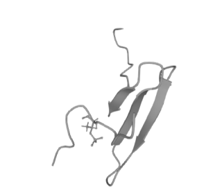
Figure 1 Schematic of the molecular structure of Tau proteins
Biological functions of P-tau proteins
Neuronal fiber tangles (NFTs) composed of highly phosphorylated tau protein (p-tau) are one of the hallmark pathological features of Alzheimer's disease (AD). p-tau181 in cerebrospinal fluid (CSF) has been recognized as a marker of NFTs and is used to support the diagnosis of clinical AD and the prediction of mild cognitive impairment. However, many phosphorylation sites of p-tau including p-tau181, p-tau217, andp-tau231 have shown their potential to be diagnostic markers for AD. The expression of some of these species of p-tau in cerebrospinal fluid does not coincide with that in plasma and brain, and the enrichment of p-tau in cerebrospinal fluid and plasma is thought to be amyloid-driven and more indicative of AD pathology.
KMD Bioscience (KMD Bioscience) has a mature in vitro diagnostic reagent development platform, all raw materials have passed multiple application platforms and repeated validation, and after strict production process control, we can continuously and stably provide high-quality raw materials for our IVD customers. Most of our antigenic raw materials are prepared using mammalian expression system, which is closer to natural antigen in structure and activity. Each antigen is produced through rigorous program design and strict performance evaluation, with excellent stability and specificity. Moreover, our antigenic raw materials can be used in the development of standards, calibrators, quality control products and as immunogens.
FL-4, Building A5, International Enterprise Community, Tianjin, China
Email:[email protected]
Hot Line: +86-022-82164980
Technical Support:[email protected]
