Service Line:+86-022-82164980
Address:FL-4, Building A5, International Enterprise Community, Tianjin, China
Email:[email protected]
KMD Bioscience has a mature antibody production platform with well-established antibody engineering technology to ensure the quality of service. KMD Bioscience has extensive experience in phage display antibody discovery and supported by our technology and platform, KMD Bioscience can provide high-quality human Fab antibody library service. KMD Bioscience is constantly upgrading its technology to better serve your needs. With many years of research and development, KMD Bioscience has established a solid platform for Fab library production. Our scientists can offer high-quality phage display library construction and custom phage display library screening services to precisely meet our clients' demands.
Fab fragments are commonly produced either enzymatically using papain to cleave intact antibodies above the hinge region or through recombinant DNA technology. Recombinant production offers the advantage of expressing Fab fragments in various host cells, including bacteria, yeast, and mammalian cells, allowing for scalability and modification flexibility. The smaller size of recombinant Fabs antibody enhances tissue penetration compared to intact antibodies. Additionally, they exhibit reduced cross-reactivity with Fc receptors on cells, resulting in decreased nonspecific binding. Moreover, Fab fragments can be humanized or fully humanized to minimize the risk of immunogenicity in therapeutic applications.
Antibody Fragment Fab
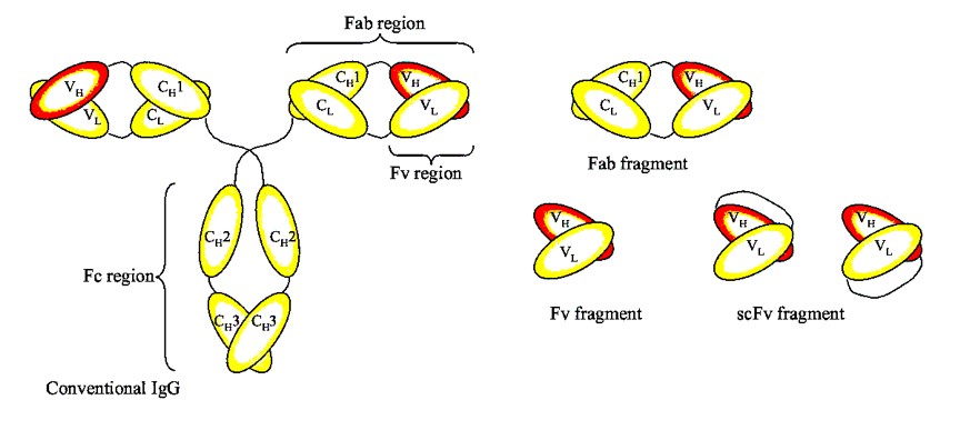
The basis for antibody recombination is the ability to obtain sequence information from monoclonal hybridoma antibodies. The Fab fragment is the main region for antibody recombination. The Fab fragment, which is the antigen binding region, determines an antibody's specificity and affinity. The Fab fragment consists of one constant (C) domain and one variable (V) domain from both the heavy (H) and light (L) chains of the antibody. These domains are connected by a disulfide bridge, and the Fab fragment includes the part of the antibody that binds to antigens. The molecular weight of a Fab fragment is approximately 50 kDa.

The Fab antibody library refers to VH and VL, especially the CDR region genes of the antibody form a large number of different clones by different strategies, such as amino acid mutations and frameshift mutations. In addition, KMD Bioscience can provide a suitable solution: after sensitizing human peripheral blood monocytes in vitro, high-affinity monocytes were isolated by flow cytometry, and then EBV monoclonalization was conducted. Using this method, highly consistent monocyte clones can be obtained, then carry out library construction.
Single chain variable fragment (scFv) and fragment antigen-binding (Fab) are engineered antibodies used in research, diagnosis and therapy applications; they differ however in structure and characteristics. scFvs are often preferred for applications requiring small size and easy genetic manipulation, while Fabs antibody are chosen for applications needing higher stability, solubility, and a longer half-life.
Type of Phage Display System
1. M13 Phage Display
Description: The M13 phage display system is one of the most widely used. It relies on filamentous bacteriophages that infect E. coli. Proteins or peptides of interest are typically fused to the minor coat protein (pIII or pVIII), allowing for the display of the fused protein on the phage's surface.
Applications: Ideal for displaying peptides and small proteins for antibody discovery, epitope mapping, and peptide library screening.
2. T7 Phage Display
Description: This system uses the lytic bacteriophage T7. The protein of interest is fused to capsid proteins on the T7 phage, such as the 10B capsid protein. Unlike filamentous phages, T7 is a lytic phage, meaning it lyses the host cell upon replication.
Applications: Suitable for displaying peptides and proteins for interaction studies and library screenings. The lytic life cycle allows for rapid and high-level display of fusion proteins.
3. λ Phage Display
Description: Lambda (λ) phage display uses the lytic bacteriophage λ. Fusion proteins can be displayed by integrating them into the D or J coat proteins. The λ system allows for larger proteins to be displayed compared to filamentous phage systems.
Applications: It's particularly useful for the display of larger protein domains or full-length proteins, making it suitable for studying protein-protein interactions and enzyme-substrate interactions.
4. T4 Phage Display system
Description: The bacteriophage T4 is a lytic phage that infects E. coli bacteria. It has a complex structure with a large head (capsid), a tail, and tail fibers. The T4 genome is relatively large compared to other phages used in display technologies, encoding for more than 300 proteins.
Applications: While the size and complexity of T4 might offer some unique opportunities for protein display, especially for large proteins or complex assemblies, its use in phage display technology is less documented in the literature compared to filamentous phages like M13 or lytic phages like T7.
KMD Bioscience has extensive experience in the phage display antibody discovery. Our phage display platform is well-established and we have been able to use it for various types of phages. The M13 phage platform is the most widely used for phage display. It is also extensively used in many research areas. There are a variety of phage display services available via T4 and T7 phage system, which may offer different promising options depending on their particular merits.
Fab Antibody Library Service Process
.jpg)
Fab Antibody Library Service Workflow
|
Steps |
Specification |
Timeline |
Deliverables |
|
Monoclonalization of peripheral blood mononuclear cells |
* Blood mononuclear cell acquisition. * Monoclonalization. * Monoclonal culture after flow cytometry. |
8-12 weeks |
* Experimental report: including detailed construction procedures and representative sequence information.
* Product delivery: including bacterial and phage antibody libraries. |
|
cDNA synthesis (Clients can provide cells) |
* Total RNA extraction. * PCR amplification with Actin-specific primers to identify the quality of cDNA. |
||
|
Library construction |
* Primer design & synthesis * PCR amplification of variable region genes of heavy and light chains using cDNA as a template. * Fab gene splicing. * Plasmid construction & transformation: after enzyme digestion, Fab and phagemid vector were ligated and transformed into TG1 host bacteria by an electric shock to construct antibody libraries. * Identification: 20-50 clones were randomly selected, PCR identification, sequencing and analysis of antibody sequences. * Packaging: >1×10^13 phage particles /ml. |
Fab Antibody Library Service Highlights
.jpg)
How to Order?

If you have any questions regarding our services or products, please feel free to contact us by E-mail: [email protected] or Tel: +86-400-621-6806