400-621-6806

Service Line:+86-022-82164980
Address:FL-4, Building A5, International Enterprise Community, Tianjin, China
Email:[email protected]
KMD Bioscience has been working on protein expression research for over 10 years. Our scientists, who have accumulated a wealth of experience in experimental operations, can quickly complete the specified experiments in a relatively short period while ensuring that the test results are accurate, objective, and credible. KMD Bioscience has established a perfect protein platform with sophisticated protein detection equipment and technology, which can provide one-stop technical services from recombinant protein production to protein purification, protein function research, and other one-stop technical services to ensure high-quality service content. We can provide customers with Western Blot analysis and other commonly used immunological detection services.
Western Blot, is one of the common technical methods for qualitative and quantitative detection of protein expression. The primary aim of Western Blotting is to detect if a certain protein (target protein) is expressed in a sample (tissue or cell) and the relative abundance of expression. Through the specific recognition and binding of antibodies to the target proteins in gel electrophoresis-treated cells or biological tissue samples, the expression of specific protein levels in cells or tissues can be analyzed according to the location and depth of coloration. This technology is a new immunochemical technology developed based on gel electrophoresis and solid-phase immunoassay technology, so it has both the high resolution and high specificity of SDS-PAGE and solid-phase immunoassay technology, which plays an important role in proteomics as a convenient and reliable research tool, and mass spectrometry and protein chip technology.
Western Blot test experimental services have developed to this day and are widely used. However getting a clear background, bright strips, and real and objective experimental results is not easy, and it takes a lot of time and effort. KMD Bioscience has the skills to summarize the details of experimental operations, has strict quality control points, and rigorous pre-experimental settings, can tailor different experimental protocols for different customers' samples, and provide clear and complete Western Blotting analysis experimental results, saving customers' valuable scientific research time and meeting customer needs with high quality.
|
Western Blot |
|
|
Experimental principle |
The technique is based on the principle of specific binding of antigen and antibody to detect a protein in complex samples. Proteins in cell or tissue samples are separated by polyacrylamide gel electrophoresis, and after membrane transfer and sealing, the target protein binds to a specific primary antibody, which reacts with a horseradish peroxidase (HRP)-labeled secondary antibody, and then undergoes substrate chromatography, which analyzes the position of the exposed bands and grayscale analysis to determine the expression of the target protein in the sample. |
|
Applications |
*Widely used for protein expression level detection. *Identification of a protein, as well as quantitative and semi-quantitative analysis of the target protein. *Can be used for subsequent analysis of protein-protein, protein-DNA, and protein-RNA interactions. |
|
Advantages |
*The required equipment is convenient, low-cost, and flexible to use. *Simple operation, generally stable results, and positive results are generally more reliable. *Results analysis is easier than mass spectrometry. |
The Main Process of the Western Blot Experiment
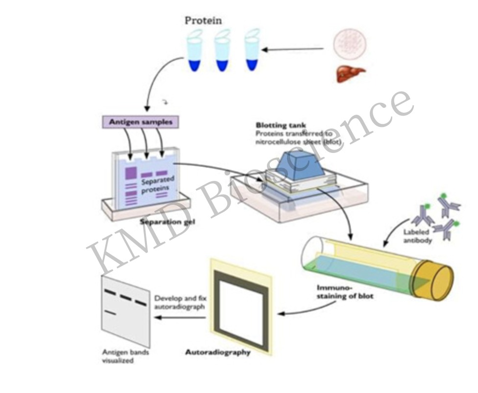
With the upgrading of electrophoresis and electrotransfer equipment, the commercialization of antibodies and signal detection systems, and other technological advances, the sensitivity of Western Blotting has been improved, and Western Blot is easy to operate and therefore can be performed in general laboratories, but a variety of problems are often encountered in the process of experimentation.
Frequently Occurring Problems and Countermeasures
|
Experimental Problems |
Possible Reasons |
Recommendations |
|
The destination strip is not visible |
Expression of the target protein is too low |
Increased sample volume, concentration of target proteins, use of higher sensitivity kits |
|
Insufficient potency of antibodies used |
Use freshly prepared primary antibodies, avoid repeated freezing and thawing; increase the concentration of primary or secondary antibodies, and prolong the incubation time appropriately. |
|
|
Low efficiency of membrane transfer |
Selection of the appropriate film transfer method to improve film transfer efficiency |
|
|
The strips are non-parallel and curved |
Electrophoretic migration too fast or migration temperature too high |
Adjust electrophoresis settings such as PH, voltage, etc., and lower the ambient temperature during electrophoresis |
|
The background is very high |
Inappropriate closure conditions |
Extend the sealing time or change the sealer |
|
High antibody concentration |
Reduced primary/secondary antibody concentration and extended incubation time |
|
|
Antibodies cross-react with sealers |
Add heat and a stain remover such as Tween 20 |
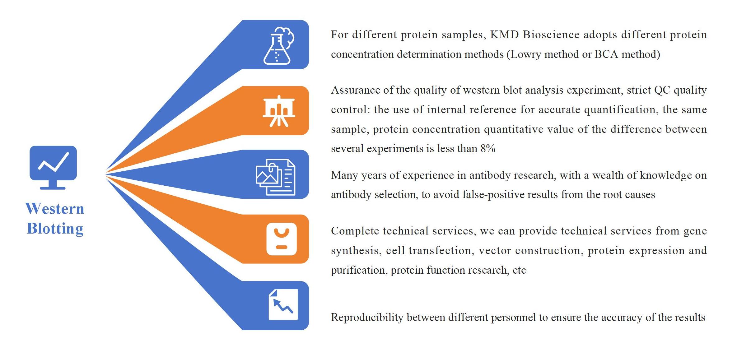
Western Blot Service Advantages
FAQ-Western Blot
F1: The destination strip is not being detected or the signal is weak.
A1: 1. Protein degradation. ------->Solution: Add a protease or phosphatase inhibitor when preparing the sample, and store it separately to avoid repeated freeze-thaw cycles.
2. Protein intake is too low. ------->Solution: A higher concentration of loading buffer can be selected to avoid protein concentration reduction while increasing the loading volume. A positive control group was established to eliminate operational problems and enhance the accuracy of protein expression verification.
3. Small proteins are lost due to long electrophoresis time. ------->Solution: Reduce the electrophoresis time or voltage.
4. Electrophoresis or film transfer instrument power supply is reversed. ------->Solution: Make sure the electrodes are properly connected through inspection.
5. PVDF membrane is not activated. ------->Solution: Activate the PVDF membrane with methanol/ethanol for 1min then set it aside.
6. Insufficient transmembrane. ------->Solution: Reformulation of transmembrane fluid; Confirm the appropriate transfer time.
7. Primary or secondary antibody. ------->Solution: Check whether the species of the primary and secondary antibodies are the same, or replace the primary antibody to avoid failure.
8. Washing film for a more or longer time. ------->Solution: Appropriately reduce the number and time of film washing.
F2: The strip is fuzzy and trailing.
A2: 1. Gel plate. ------->Solution: Gel formulated with unclean or undried gel plates is prone to bubbles.
2. Sample delivery. ------->Solution: The protein sample is fully dissolved to prevent absorption, precipitation, and cross-contamination.
3. Transmembrane. ------->Solution: The bubble is not removed or the "sandwich" is not tightened when the film is transferred resulting in the film is not sufficient.
F3: The background is too high.
A3: 1. Inadequate closure. ------->Solution: Extend the sealing time or increase the concentration of the sealing liquid.
2. Washing is not sufficient. ------->Solution: Increase the time and frequency of washing to remove non-specific binding.
3. The exposure time is too long. ------->Solution: Reduce exposure time.
F4: Multiple strips appear or the positions of the strips are incorrect.
A4: 1. Inadequate closure. ------->Solution: Extend the sealing time or increase the concentration of the sealing liquid.
2. The specificity of the primary antibody is poor. ------->Solution: Replace the primary antibody.
3. The concentration of the secondary antibody is higher. ------->Solution: Increase the dilution ratio and reduce the antibody dosage of the secondary antibodies.
4. There are splinters in the target protein. ------->Solution: In the presence of splinters, the digestion of cells was reduced before protein extraction, and sequences and sites were queried by BLAST.
F5: The strips are uneven.
A5: 1. The problem of making glue. ------->Solution: Select and make the appropriate concentration of glue, and pull out the comb smoothly after the glue sets, which prevents the strips from arching due to the convex point sample hole surface.
2. The amount of protein sample. ------->Solution: Reduce the amount of protein in the sample to avoid the sample appearing in other holes.
3. The electrophoresis speed is too fast. ------->Solution: Reduce the electrophoresis speed to ensure that proteins can be adequately isolated and localized to avoid the "smile" strip.
4. Electrophoresis temperature is too high. ------->Solution: Adjust the temperature of the electrophoresis system to avoid the phenomenon of the fuzzy strip, because the temperature is too high that can’t accurately separate proteins of different sizes.
How to Order?

If you have any questions regarding our services or products, please feel free to contact us by E-mail: [email protected] or Tel: +86-022-8216-4980;
FL-4, Building A5, International Enterprise Community, Tianjin, China
Email:[email protected]
Hot Line: +86-022-82164980
Technical Support:[email protected]
