400-621-6806

Service Line:+86-022-82164980
Address:FL-4, Building A5, International Enterprise Community, Tianjin, China
Email:[email protected]
KMD Bioscience has been committed to cell biology technology research for many years, cells are the basic structural and functional units of life activities, and cell-level experimental techniques play a vital role in research in the field of life sciences. KMD Bioscience has established a complete cell culture platform that can complete a variety of basic cytology experiments, and we are able to provide customers with cell scratch experiment services. KMD Bioscience has an experimental team composed of experienced researchers, our scientists have accumulated rich experience in cell technology research, and have a unique summary in each cell experiment operation step, which can quickly complete the specified experiment in a very short time, with mature technology, obtain high-quality "scratches", ensure the consistency of "scratches", and have good experimental repeatability to ensure that customers get ideal and reliable experimental results, which can not only shorten the experimental cycle, but also save valuable scientific research time. In addition, KMD Bioscience has built a complete cell biology technology service platform, relying on advanced immunofluorescence microscopy and other technical equipment, which can provide customers with high-quality imaging technology, provide high-definition photo results, and improve customer experience.
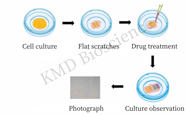
Cell scratch assay is a simple, convenient and economical in vitro test method to study cell migration ability, which can be used to detect the invasion and metastasis ability of growing tumor cells. Cell scratch experiment to study cell motility characteristics, similar to in vitro wound healing model, in vitro dish or plate culture of monolayer adherent cells, with a small amount of tip or other hard objects in the central area of cell growth, remove the central part of the cells, and then continue to culture the cells to the time set by the experiment, take out the cell culture plate, observe the growth and migration ability of surrounding cells, the experiment usually needs to set the normal control group and the experimental group, the experimental group is a group with a certain treatment factor or drug, exogenous genes, etc. The migration and repair ability of cells in each group can be judged by the difference in the repair ability of cells in the scratch area between different groups. Basic steps include creating a "wound" in the cell monolayer, capturing images at the beginning of the cell migration process and periodically determining the cell migration rate by comparing the images. To a certain extent, the scratch experiment simulates the process of cell migration in vivo and is a relatively simple method among the in vitro test methods for studying cell migration.
|
Project |
Cell Scratch Experiment Services |
|
Experimental principle |
* The basic principle of the technology is: the cells are cultured on a petri dish or plate when the cells grow to a monolayer state, an artificial blank area is created on the fused monolayer cells, that is, a "scratch", and the cells on the edge of the scratch will gradually enter the blank area under appropriate conditions to heal the "scratch"; By observing the cell state of the scratched area at different times, the migration ability of cells can be judged |
|
peculiarity |
* It has the advantages of low experimental cost, convenient operation and economic efficiency
* Suitable for studying cell migration caused by cell-to-extracellular matrix (ECM), cell-to-cell interaction
* Compatible with microscopy systems including live-cell imaging for analysis of cell-cell interactions |
|
apply |
* This method can mimic the ability of cells to migrate in the body
* It can also be used to verify whether the proliferation rate and migration ability of cells change after different substances stimulate cultured cells |
|
Experimental Steps |
Cycle |
||
|
Sample Delivery Requirements |
Please contact our customer service before sending samples |
|
|
|
Experimental Steps |
(1) All experimental instruments should be sterilized |
2-3 weeks |
|
|
|
(2) Draw a line evenly behind the cell culture plate |
|
|
|
|
(3) Add an appropriate amount of cells to each well so that it can be spread overnight |
|
|
|
|
(4) The next day, scratch the tip perpendicular to the horizontal line on the back of the plate, and the tip should be vertical |
|
|
|
|
(5) Wash three times with PBS, wash off the streaked cells, and add serum-free medium |
|
|
|
|
(6) Put it in the incubator and take it out at regular intervals to take pictures |
|
|
|
Deliverable: Cell scratch photos, detailed lab reports |
|
||
Service Advantages:
--Completely solve cell scratch experiments, from sample preparation to image analysis in just a few steps
--High throughput enables effective comparisons between samples and reduces the impact of batch-to-batch variation
--Advanced cell laboratory, complete cell culture platform, scientific experimental group and control group comparison design, to ensure that the data results are objective, authentic and credible
--Perfect experimental operation level, perfect experimental conditions, strict cell experiment control system, experimenters have many years of successful experience in cell experiments, providing better repeatability of experimental results
--Provide detailed and complete experimental reports, original experimental results, including pictures before and after experiments, and provide clear electronic pictures and analysis results of cell scratch experiments
--Complete instrument platform: with multi-type microscope, flow cytometer, enzyme-linked immunodetector and other important instruments, high sensitivity, wide linear range, good experimental repeatability
--Provide one-stop technical services from cell culture, cell staining to picture photography, data analysis and so on
How to Order?

If you are interested in our service offerings, please directly call the hotline at+86-400-621-6806 or send an email to [email protected]
FL-4, Building A5, International Enterprise Community, Tianjin, China
Email:[email protected]
Hot Line: +86-022-82164980
Technical Support:[email protected]
