400-621-6806
Service Line:+86-022-82164980
Address:FL-4, Building A5, International Enterprise Community, Tianjin, China
Email:[email protected]
Influenza is a disease caused by influenza viruses. Influenza viruses are categorized as type A, B and C based on different nucleolar and matrix proteins. Influenza viruses are categorized as type A, B, or C based on complement-binding antibodies to nuclear and matrix proteins. Hemagglutinin (H) is a glycoprotein on the surface of influenza viruses that allows the virus to bind to cellular salivary acids and thus fuse with host cell membranes. Neuraminidase (NA) is another surface glycoprotein that removes salivary acid to prevent the release of the virus from the infected host.
Influenza A virus (FluA) is a common influenza virus, Influenza A virus is most prone to mutation, and human avian influenza (referred to as human avian influenza) is an acute respiratory infectious disease caused by some strains of certain subtypes of avian influenza A viruses, viral gene mutation can infect humans, the symptoms of the infection mainly manifested as high fever, coughing, runny nose, myalgia, etc., most of which are accompanied by severe pneumonia. The symptoms of infection mainly include high fever, cough, runny nose, myalgia, etc. Most of them are accompanied by severe pneumonia, and in severe cases, heart, kidney and other organs fail, leading to death, and the case fatality rate is very high.
Influenza B virus (FluB), is caused by influenza B (B) virus caused by influenza, which is characterized by a rapid onset of illness, chills, fever, body temperature in a few hours to 24 hours to reach the peak, 39-40 ℃ or even higher.
KMD Bioscience, as a supplier of biological raw materials, develops antigens and antibodies covering the fields of infectious diseases, fertility, cardiac markers, tumor markers, inflammation and serum proteins, which are widely used in various technology platforms, such as immunochromatography, ELISA, chemiluminescence and PCR. After years of innovation and development, the company has built several technology platforms, including colloidal gold/latex immunochromatography technology platform, fluorescence immunology technology platform, chemiluminescence technology platform and molecular diagnostics technology platform.
The inventory of reagents associated with FLUA and FLUB that KMD Bioscience can offer:
|
CAT# |
Product Name |
Species |
Host |
Application |
Size |
Inquiry |
|
VAG362 |
Virus |
Vero cell |
ELISA |
1ml |
Inquiry |
|
|
KMP2204 |
|
|
|
1ml |
Inquiry |
|
|
PA278 |
Human |
Mouse |
LFIA (Lateral-Flow Immunochromatographic Assay), CLIA (Chemiluminescence Immunoassay), ELISA |
1mg |
Inquiry |
|
|
PA279 |
Human |
Mouse |
LFIA (Lateral-Flow Immunochromatographic Assay), CLIA (Chemiluminescence Immunoassay), ELISA |
1mg |
Inquiry |
|
|
PA280 |
Human |
Mouse |
LFIA (Lateral-Flow Immunochromatographic Assay), CLIA (Chemiluminescence Immunoassay), ELISA |
1mg |
Inquiry |
|
|
PA281 |
Human |
Mouse |
LFIA (Lateral-Flow Immunochromatographic Assay), CLIA (Chemiluminescence Immunoassay), ELISA |
1mg |
Inquiry |
Molecular structure and subtypes of FluA and FluB
Influenza A viruses are divided into many subtypes according to H and N antigens, with 18 subtypes of H (H1 to H18) and 11 subtypes of N (N1 to N11). Among them, only H1N1, H2N2, H3N2 mainly infect humans, H1N1 and H3N2 are the main subtypes that cause seasonal influenza, and the natural hosts of many other subtypes are a variety of birds and animals.
Influenza A viruses are most prone to mutation, and influenza pandemics are caused by the emergence of new subtypes of influenza A viruses or the reemergence of old subtypes. The surface antigen of influenza A viruses often undergoes small mutations, which are called "antigenic drift" (antigenicdrift). Figuratively speaking, "drift" means that the virus camouflages itself through small changes to avoid recognition by the human immune system. The human immune system to avoid the recognition of the purpose. The result of antigenicdrift is that the strain of influenza that causes influenza may be different each year, and people will need to be revaccinated against influenza each year.
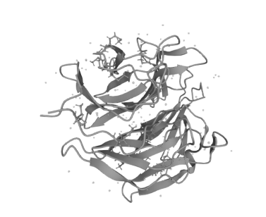
Figure 1 Schematic structure of FluNS1A-molecule
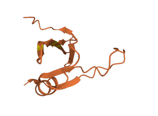
Figure 2 Schematic diagram of the molecular structure of FLUB_ASPFN.
Biological characteristics of FluA and FluB
Influenza viruses usually rely on the binding of certain parts of viral proteins to specific proteins in the human body to invade the human body, because through such binding, influenza viruses are able to inhibit the body's own natural defenses against viral infections, paving the way for effective replication of the viruses in the human body.
Antigenic shift occurs only with the A virus. It occurs when the same cell is infected with 2 viruses, human and animal, and genetic reassignment occurs between the viruses. The resulting viral hemagglutinin and neuraminidase undergo a new binding, leaving the population without immunity. The antigenic shift is responsible for the global pandemic of influenza. Influenza A viruses undergo a major mutation approximately every decade or so, and since 1933 the A viruses have undergone 4 antigenic shifts: H0N1 (original A, A0) from 1933-1946, H1N1 (subtype A, A1) from 1946-1957, H2N2 (Asian A, A2) from 1957-1968, and after 1968 H3N2 (Hong Kong type, A3). There is generally a marked alternation between the old and new subtypes, with the old subtypes no longer being isolated after the new subtype has emerged and become endemic to an area. The same major and minor variants are found between influenza B infections, but they are not divided into subtype shifts. No antigenic variation has been detected in influenza C viruses.
KMD Bioscience, with years of experience in R&D and advanced technology platforms, launches high-quality "all-round" technology services, including antibody discovery (hybridoma monoclonal antibody technology, Beacon single-cell sorting technology, phage display technology), protein expression (prokaryote expression, yeast protein expression, phage display technology), and antibody discovery. , yeast cell protein expression, insect cell protein expression, mammalian cell protein expression). Our antigens are produced after rigorous program design and performance evaluation, and have excellent stability and specificity. In addition, our antigen materials can be used in the development of standards, calibrators, quality control products and as immunogens.
FL-4, Building A5, International Enterprise Community, Tianjin, China
Email:[email protected]
Hot Line: +86-022-82164980
Technical Support:[email protected]
